3d brain mri classification
For the sake of simplicity this data was named dataset-I in this work This dataset contained a total of 3000 brain MRI images of. The model called VoxResNet consists of volumetric residual blocks VoxRes blocks containing convolutional layers as well as several.

Brain Mri In A 55 Year Old Male Fabry Patient Gla Variant P Download Scientific Diagram
Implement 3d_DenseNet_mri with how-to QA fixes code snippets.
. A One axial slice of the segmented brain. Pyradiomics a python open-source package is used to extract GLCM features. The elaboration of this new module its labeling of more than 524 structures on 379 MRI images in three different.
In this work three different online datasets of brain MRI images were utilized. The best performing model is chosen among different classifiers. The detailed anatomical information of the brain provided by 3D magnetic resonance imaging MRI enables various neuroscience research.
MRI Dataset for Brain Analysis The data evaluation framework of three-dimension 3D cross-sectional brain MRI is used to classify patients with AD and to segment brain tissue types CSF GM and WM. D Result after morphological operations. The 3D model reached 93 on the training set and 93 on the test set.
For example for brain segmentation an architecture similar to ResNet was proposed which expands the possibilities of deep residual learning for processing volumetric MRI data using 3D filters in convolutional layers. Proper treatment planning and accurate diagnostics should be implemented to improve the life expectancy of the patients. 2017 IEEE 14th International Symposium on Biomedical Imaging ISBI 2017 Article.
Finally we conclude with a general discussion and explore future directions in the field of brain MRI segmentation. Anatomy of the brain MRI - cross-sectional atlas of human anatomy. 3d_DenseNet_mri Code for 3d_DenseNet for 3D Brain MRI Classification Machine Learning library by newcolour1994 Python Version.
Kandi ratings - Low support No Bugs No Vulnerabilities. The module on the anatomy of the brain based on MRI with axial slices was redesigned having received multiple requests from users for coronal and sagittal slices. B Result of FPCM classification.
These radiomics features can uncover the characteristics of the disease. Spatiospatial were employed to classify brain tumours of the different types of gliomas based on their grading as well as healthy brains from 3D volumetric MR Images using a single MR contrast. We used the morphometry data for gender classification with machine learning models in comparison as one of the most popular methods for data analysis in neuroimaging.
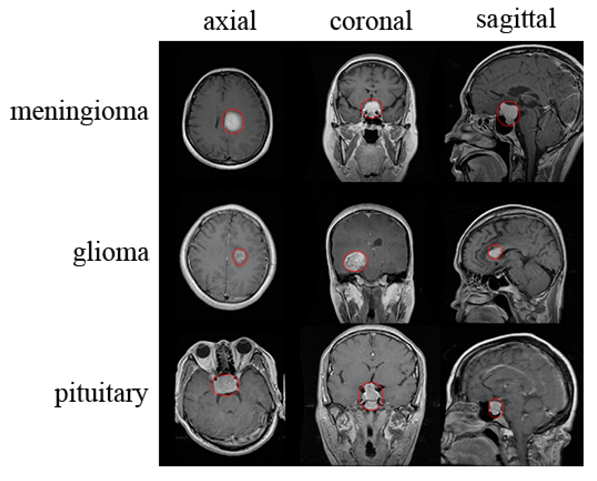
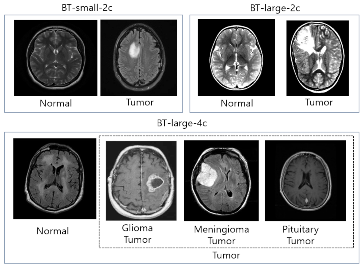
In this paper 3D MRI is employed for the detection and classification of a Brain Tumor. Benign Tumor Malignant Tumor Pituitary Tumor etc. Fayad Christoph Lippert Conference paper First Online.
We demonstrate the performance of the proposed approach for classification of Alzheimers disease versus mild cognitive impairment and normal controls on the Alzheimers Disease National Initiative ADNI dataset of 3D structural MRI brain scans. Magnetic Resonance Imaging MRI Dataset. The 3D model used 80 slices from 150 slices 35 to 115 the 2D models with 3 slices slices 747576 and 1 slice from 150 slices.
45 forks Releases No releases published. A huge amount of image data is generated through the scans. Results obtained in the classification step for two 3D images.
Radiomics uses data-characterization algorithms capable of getting a large number of features from MRI. However due to the long scan time for 3D MR. Taking into account the importance of detection of brain tumor this paper analyzes four architectures of convolutional neural networks CNN for classification of brain MR images into tumorous or.
To examine the feasibility of applying CNN to classification of schizophrenia and controls based on structural Magnetic Resonance Imaging MRI we built 3D CNN models with different architectures and compared their performance with a handcrafted feature-based machine learning approach. Submitted on 23 Jan 2017 Residual and Plain Convolutional Neural Networks for 3D Brain MRI Classification Sergey Korolev Amir Safiullin Mikhail Belyaev Yulia Dodonova In the recent years there have been a number of studies that applied deep learning algorithms to neuroimaging data. Deep Learning Models for 3D MRI Brain Classification A Multi-sequence Comparison Marius Pullig Benjamin Bergner Amish Doshi Anja Hennemuth Zahi A.
The best technique to detect brain tumors is Magnetic Resonance Imaging MRI. 3D brain tumor segmentation in MRI using fuzzy classification symmetry analysis and spatially constrained deformable models. XGBoost k-Nearest Neighbors KNN and Logistic Regression LR with a grid-search.
Code for Residual and Plain Convolutional Neural Networks for 3D Brain MRI Classification paper neuroml. Brain Tumors are classified as. This interactive brain model is powered by the Wellcome Trust and developed by Matt Wimsatt and Jack Simpson.
Reviewed by John Morrison Patrick Hof and Edward Lein. No packages published. 05 April 2022 235 Accesses Part of the Informatik aktuell book series INFORMAT Zusammenfassung.
C Selected tumor class. Structure descriptions were written by Levi Gadye and Alexis Wnuk and Jane Roskams. No License by newcolour1994 Python.
The first publicly available dataset of binary-class brain MRI images was downloaded from the Kaggle website. No License Build not available.
Brain Tumor Images A Magnetic Resonance Imaging In Mri The Magnetic Download Scientific Diagram
Diagnostics Free Full Text Brain Tumor Detection And Classification On Mr Images By A Deep Wavelet Auto Encoder Model Html
Brain Lesion Detection In Mri Images With Graph Cut Algorithms

Classification Of Brain Tumours In Mr Images Using Deep Spatiospatial Models Scientific Reports

Example Of Different Brain Mri Images Presenting Different Alzheimer S Download Scientific Diagram

Evaluation Of The Intra And Inter Method Agreement Of Brain Mri Segmentation Software Packages A Comparison Between Spm12 And Freesurfer V6 0 Physica Medica European Journal Of Medical Physics

Different Types Of Artifacts In Brain Mri A Chemical Shift B Download Scientific Diagram

Sample Datasets Of Brain Tumor Mri Images Normal Brain Mri 1 To 4 Download Scientific Diagram
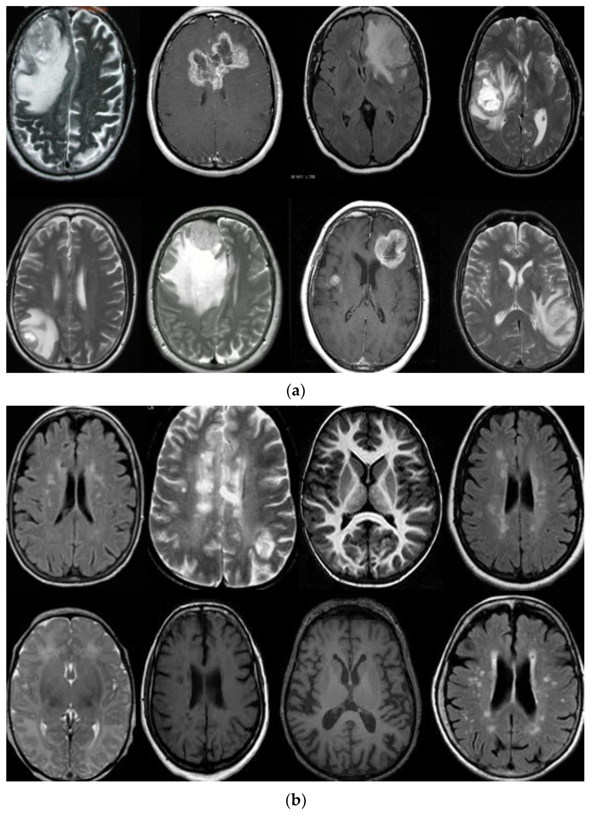
Applied Sciences Free Full Text Classification Of Brain Tumors From Mri Images Using A Convolutional Neural Network Html
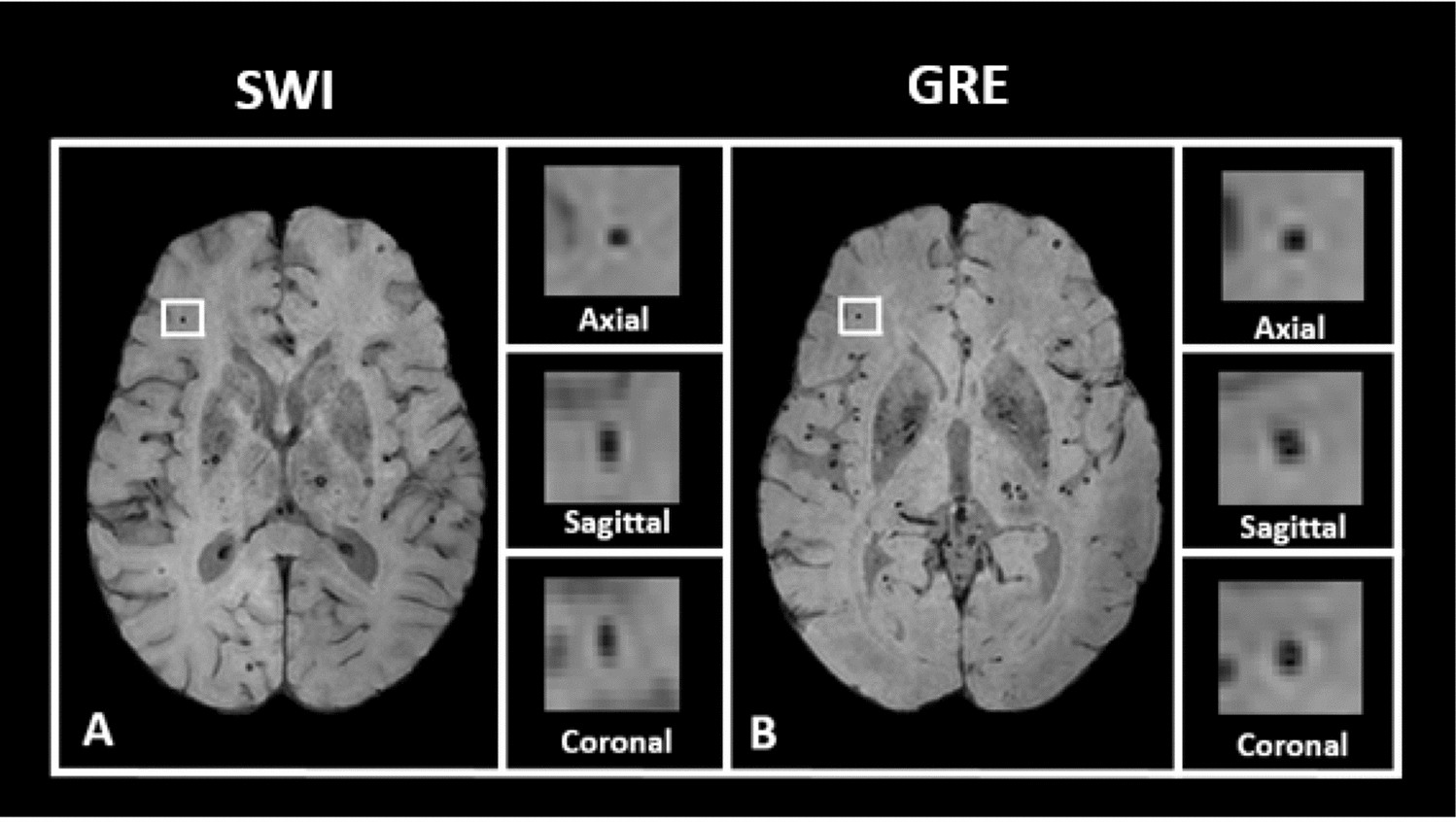
Automated Detection Of Cerebral Microbleeds On T2 Weighted Mri Scientific Reports

Brain Mri Obtained From A Sagittal Plane B Axial Plane And C Download Scientific Diagram
Diagnostics Free Full Text Mr Imaging Of Pediatric Brain Tumors Html
Classification Of Brain Tumours In Mr Images Using Deep Spatiospatial Models Scientific Reports

Fwnnet Presentation Of A New Classifier Of Brain Tumor Diagnosis Based On Fuzzy Logic And The Wavelet Based Neur Learning Methods Machine Learning Fuzzy Logic

Brain Mri Obtained From A Sagittal Plane B Axial Plane And C Download Scientific Diagram

Brain Mri Age Classification Using Deep Learning File Exchange Matlab Central

Standardized Brain Mri Protocol To Evaluate Patients In Whom Multiple Download Scientific Diagram
Sensors Free Full Text Mri Based Brain Tumor Classification Using Ensemble Of Deep Features And Machine Learning Classifiers Html

Different Angles Of An Mri A Sagittal B Axial And C Coronal Download Scientific Diagram